What is a macular hole?
A macular hole is a small break in the macula, the part of your eye responsible for detailed, central vision.
Although the symptoms can be very similar to age-related macular degeneration (AMD), the condition is very different.
It’s not clear why some people develop a macular hole and others don’t.
The vitreous is a jelly-like substance that fills up the space inside your eyeball. It changes as you age. It becomes more watery and usually moves away from the back of the eye (where it is initially attached to the retina) towards the centre of your eye.
Usually, these changes to the vitreous don’t cause problems to your vision. You may notice an increase in floaters or flashes in your vision. Floaters usually appear as a small darkish fleck (like a strand of cotton, or a little spider web) that moves around in your field of vision.
In some people the vitreous jelly is firmly attached to the retina at the macula. As the vitreous shrinks, it can pull on the macula, causing a small tear. This is the start of a macular hole. Fluid can then seep into the hole, causing sight to become blurred and distorted.
Over time, the hole can get bigger, causing more vision problems.
If your optometrist finds or suspects you have a macular hole, you may be referred to an ophthalmologist.
Macular holes usually only affect one eye, although there is a 15 to 20 per cent chance that the other eye will also get a hole at some stage.
Symptoms of a macular hole
A macular hole causes changes in the central part of your vision.
These changes can range from straight lines looking wavy in the early stages to a small blank patch in the centre of your vision in the late stages of macular hole development.
You may first notice that you have trouble reading small print or that there is distortion when you look at a printed page.
What are floaters?
As mentioned above, changes in the vitreous may increase the number of floaters or flashes in your vision. Floaters usually appear as a small darkish fleck (like a strand of cotton, or a little spider web) that moves a little around your field of vision. If you notice a significant increase in new floaters or flashes it is important to make an appointment with your eye care professional – optometrist or ophthalmologist.
Stages of a macular hole
There are a number of different stages to a macular hole. These stages are usually classed by the size of the hole and the layers of the eye that are affected. This is important to know because in the very early stages it’s possible that a macular hole may heal or spontaneously resolve without any treatment.
This means that sometimes your ophthalmologist will simply want to regularly monitor a very small hole before deciding whether or not to treat it.
However, in most cases, a macular hole will get bigger and distort vision, so treatment may be needed. Treatment attempts to stop the hole developing to a stage where most of your central vision can be lost.
Preventing a macular hole
There’s nothing you can do to prevent a macular hole. Diet or exercise are not thought to contribute to the problem.
There’s no evidence that taking any kind of medicine or vitamins can help fix a macular hole. In most cases the best treatment is surgery.
Having an eye test at least every two years is the best way to help ensure that any eye issues are detected early. In between it is important to monitor your own vision with the weekly use of an Amsler Grid.
Treating a macular hole
Most macular holes require surgery.
Your ophthalmologist will normally prefer to operate on a macular hole within a month or two of it being found. The longer a hole is left, the larger it will normally become and the harder it is to successfully close the hole.
In most cases, surgery will stop your vision problems getting worse. Most people will notice some improvement in vision, and in more than 50 per cent of cases, patients will gain sufficient vision to allow driving and reading. However, it’s uncommon that ‘perfect’ vision is restored even when a macular hole is successfully closed with surgery.
There are two main stages to the treatment:
- surgery to remove the vitreous and insert gas into the eye
- a recovery period when the gas pushes the retina back into place and the hole closes.
Surgery for a macular hole
Macular hole surgery is generally performed under local anaesthetic.
The ophthalmologist removes most of the vitreous jelly in your eye, leaving a space into which a gas is inserted. The ophthalmologist will also normally peel away a fine membrane across the back of the eye around the macular hole.
Gas is inserted into your eye which helps the edges of the macular hole to close together. After a period (usually two to six weeks), the gas is gradually absorbed by the body and is replaced with the natural fluid made by the eye.
Recovery
Over 90 per cent of holes will close after surgery. While the gas is in place it’s normal for your vision to be poor. When this gas has been absorbed and fluid has taken its place, your sight should improve.
For many people, it can take several months for some improvement to occur. However, in others, the operation’s main effect is to stop the sight becoming any worse.
It’s uncommon to have a macular hole in both eyes, so even in the rare cases where the hole doesn’t close, most people have good vision in their other eye.
Posturing
Until recently, most people who had macular hole surgery were required to spend a significant period after the operation with their head facing downwards, to ensure that the gas bubble maintained contact with the retina.
This was a key part of recovery and is known as posturing. Posturing is now becoming increasingly unnecessary, however there may be some situations where it’s still needed. Check with your ophthalmologist if you need to posture, and if so, for how long.
If posturing is necessary, you’ll need to plan for this before the operation. You’ll most likely need some help after your procedure as well. Staying face down for several days can be hard and may be made more difficult if you have other problems such as arthritis. It’s important to discuss any other medical problems that may affect your ability to posture with your ophthalmologist.
Prepare for posturing before surgery
Preparing before you go into hospital is important. If you need to posture, you’ll be expected to start as soon as you return home. Before you go into hospital consider things such as:
- doing laundry and housework, and ensuring the home is clear of trip hazards
- paying all household bills due during your recovery time
- shopping and food preparation (e.g. prepare meals ahead and freeze)
- arranging delivery of meals or other social services
- talking to your ophthalmologist about renting posturing equipment. It may take a week or so for these to be delivered, so be sure to leave enough time
- make sure your posturing furniture or aids are positioned where you want to sit
- arranging for someone to stay with you if you live alone.

While posturing you may wish to:
- keep the things you use frequently close by (for example, tissues, drinks, books, phone, tablets, laptop)
- use a straw to help with drinking
- use a laptop or a tablet such as an iPad, instead of watching TV.
If you do need to posture, you’ll usually need to spend 50 minutes out of every hour face down. Time off from posturing is generally allowed for eating, using the bathroom and applying post-surgery eye drops.
It’s not necessary to lie completely flat. Many people can maintain the correct position by sitting in a chair. Trying out different posturing positions can help avoid stiffness and boredom. For example:
- sitting at a table and putting your head down onto the table
- lying on your side in bed with pillows propped on either side of you.
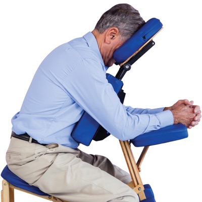
You may also consider hiring a posturing chair, which is ergonomically designed to allow you to posture without any strain on your back, chest or neck.
This chair will allow you to read, write, drink and socialise with minimal movement, while maintaining the correct posturing position. Contact MDFA for further information.
National Helpline
1800 111 709Complications of macular hole surgery
There are two main possible complications associated with the operation.
Cataract
Cataract development is considered more of a consequence of macular hole surgery, rather than a complication as such. A cataract is a clouding of the lens inside the eye. If you haven’t previously had a cataract removed from the eye that has the macular hole, it’s almost certain that a cataract will form in the months or years after macular hole surgery.
It’s worth noting, however, that everyone eventually develops a cataracts with age, even if they don’t have a macular hole. Macular hole surgery may just make a cataract form earlier. The cataract can usually be removed once it starts to affect vision. If you already have a cataract forming, many surgeons will perform macular hole surgery and cataract surgery at the same time.
Retinal detachment
When your eye surgeon removes the vitreous jelly or peels the membrane from the retina, there is a small chance that the retina may detach from the back of the eye. If this happens then your ophthalmologist will take steps to reattach the retina as soon as possible, sometimes during the same surgery.
Talk to your ophthalmologist about these and other possible complications.
Managing vision loss
Macular hole surgery normally helps maintain good vision. Rarely, a second operation may be needed to help close the hole. If surgery is unsuccessful, central vision is usually lost, as it would be if your macular hole remained untreated.
However, peripheral (side) vision will remain normal. If sight in the other eye is still good, most people adjust quite quickly and can maintain their usual normal activities.
If vision in both eyes is poor, you may need extra help. A key priority in managing vision loss is maintaining quality of life and independence Macular Disease Foundation Australia can help with information, advice and support to live well with vision loss.
Get the fact sheet
You can download a fact sheet with the information about macular hole, or head to our Resources page and you can order a free copy to be sent to you in the post.
Download the publication today.
Download


