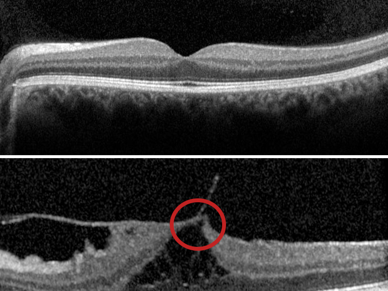
What is vitreomacular traction syndrome?
Vitreomacular traction syndrome occurs when the clear, jelly-like substance inside the eye, called the vitreous, pulls on the macula, distorting its normal shape.
This pulling isn’t painful. But because the macula is responsible for detailed central vision, it can cause your vision to become blurry and/or distorted. It may also cause straight lines to appear wavy or bent. They are similar symptoms to those experienced in other macular conditions, including age-related macular degeneration (AMD).
If you’ve been diagnosed with vitreomacular traction syndrome, it’s important to monitor your vision carefully using an Amsler grid. Consult your eye health professional immediately if you notice any sudden changes in your vision.
If there are any changes in your vision, prompt action could save your sight.

How does vitreomacular traction syndrome occur?
When we are born, the vitreous is firmly attached to the retina.
As we age, the vitreous becomes more watery. It may eventually separate from the retina in a process known as posterior vitreous detachment.
If the vitreous doesn’t fully separate from the retina, the remaining partially attached vitreous can pull and distort the macula. This is vitreomacular traction. In some cases, vitreomacular traction can progress and form a macular hole.
Diagnosing vitreomacular traction syndrome
Your optometrist or ophthalmologist can diagnose vitreomacular traction syndrome during an eye examination. An optometrist may monitor your condition but they will refer you to an ophthalmologist for any necessary treatment.
Your eye health professional may also use optical coherence tomography (OCT) to confirm the extent of your condition. OCT is a non-invasive scan of the tissue layers of the retina.
Management and treatment
There are currently three main options for managing vitreomacular traction syndrome
Observation
If symptoms don’t impact on your daily life, your ophthalmologist may decide to monitor your condition. Some cases of vitreomacular traction syndrome resolve spontaneously.
Vitrectomy
A vitrectomy is a surgical procedure where the vitreous is removed from the eye to relieve the pulling, or traction, on the macula.
The surgical procedure is usually performed under local anaesthesia.
In most cases, patients experience a significant improvement in vision. After the surgery, you’ll use special eye drops for several weeks. Your ophthalmologist will discuss the benefits and risks of surgery and provide instructions for postoperative care.
Ocriplasmin
Ocriplasmin, marketed under the brand name Jetrea, is a drug treatment administer as a single injection into the eye. It works by dissolving the adhesion between the vitreous and the macula. This, in turn, relieves the traction. Ocriplasmin is suitable only in very select cases. Your ophthalmologist will discuss whether this treatment is appropriate in your case, and the risks and benefits.
Monitoring your vision
If you’ve been diagnosed with vitreomacular traction syndrome, it’s important to monitor your vision carefully at home using an Amsler grid.
Your FREE Amsler grid
Get the fact sheet
The information on this page is available as a fact sheet for download.
Download the publication today.
Download


