What is a retinal detachment?
Retinal detachment occurs when the retina separates from the back of the eye. The retina needs to be attached to the back of the eye to survive and work properly.
A retinal detachment is an emergency and needs to be assessed and treated as soon as possible.
If a retinal detachment is not detected and treated it may result in the permanent loss of some or all the vision in your eye.
Most retinal detachments happen because a tear or hole in the retina allows fluid to accumulate under the retina. This causes the retina to detach. Holes and tears in the retina can occur when the vitreous gel that fills the middle of the eye becomes detached from the retina (known as acute posterior vitreous detachment or PVD) or due to a significant eye injury. These are known as rhegmatogenous retinal detachments.
Other eye conditions, such as diabetic retinopathy, can cause scar tissue formation, which pulls on the retina (traction) causing a tractional retinal detachment. A retinal detachment can also occur when fluid leaks under the retinal layers without a hole or tear being present. This is known as a serous retinal detachment.
Who’s at risk of retinal detachment?
You have an increased risk of retinal detachment if you are over the age of 50, have a family history of retinal detachment or you are highly short sighted.
You’re also at risk if you’ve had:
- previous eye surgery, for example, cataract removal
- trauma directly to the eye
- history of retinal detachment
- previous peripheral retinal degeneration or tears.
Symptoms of retinal detachment
Floaters
Floaters can take lots of different forms, shapes, and sizes. You may see them as dots, circles, lines, clouds, or cobwebs in your vision. Sometimes, floaters can move around quickly. At other times it can feel like they hardly move at all. You may find floaters are more obvious in bright light or on a sunny day.
A floater is created when harmless clumps of cells develop and float in the watery vitreous. These clumps cast a shadow on the retina. We see these shadows as floaters. A change in your normal floaters could be a sign of retinal detachment.
Flashes of light
Flashes of light occur when the vitreous pulls away from the retina, tugging on it. The retina reacts by sending a small electrical signal to your brain. You see this as short, small, flashes of light. Flashes can also occur when you do not have retinal detachment.
Dark shadow or grey curtain
A dark shadow or grey curtain can appear in your side vision when your retina detaches. This shadow may move towards the centre of your vision with time.
If you have any of the above symptoms – or any other visual symptoms you have not experienced before – you must get your eyes examined as soon as possible.
You can find an optometrist near you using our online directory. In case of emergency, you can also go to your nearest eye hospital, or emergency department of your local hospital.
Diagnosis
A retinal detachment is diagnosed by a comprehensive eye examination with an optometrist or ophthalmologist. Your eye health professional may dilate (enlarge) your pupils using eye drops to examine the retina at the back of your eye. After your pupils have been dilated, it is normal for your eyes to be blurry and sensitive to light for a few hours. You shouldn’t drive while your eyes are still dilated.
Prevention
Regular eye tests are an important way to monitor your eye health.
One of the causes of retinal detachment is trauma to the eye. Wearing eye protection when using tools, gardening or for sport is something you can do to reduce the risk of an eye injury.
If you experience symptoms of flashes and floaters, and your eye health professional detects a tear in your retina, this may be treated to reduce the risk of a retinal detachment developing. Not all retinal tears or holes need treating. Treatment of retinal tears or holes may be done by using a laser or a cryoprobe (“cryo”) to seal the retina and prevent fluid passing through to cause a detachment.
Treatment for retinal detachment
Treatment for rhegmatogenous retinal detachment involves surgery to reposition the retina against the back of the eye. The sooner treatment is carried out, the better the results are likely to be. If retinal detachment is not treated then you are likely to lose all the vision in the affected eye over time. A retinal detachment will not get better without treatment.
The type of surgery or combination of surgeries depends on the type and location of the detachment and any complicating factors, such as other eye conditions you may have. You should discuss the risks and benefits of surgery with your ophthalmologist.
Pneumatic retinopexy (gas bubble surgery)
A pneumatic retinopexy involves injecting a gas bubble into your eye. This bubble then presses the retina back in place, and cryotherapy or laser is applied around the hole or tear. The gas is reabsorbed over time and is replaced by fluid as the eye heals.
Vitrectomy
A vitrectomy involves removing the vitreous gel from the eye. The vitreous is replaced with a gas bubble or silicone oil which holds the retina in place against the inside of your eye. If a silicone oil is used, it is removed a few months later by the ophthalmologist.
Scleral buckle
A scleral buckle involves attaching a tiny piece of silicone material to the outside of your eye. This pushes the outside of the eye against the detached retina into a position which helps the retina to re-attach. Cryotherapy or laser treatment is used to seal the area around the retinal tear. The buckle usually isn’t removed and it’s not visible after surgery.
Recovery from retinal detachment surgery
After surgery, the eye will feel uncomfortable, possibly for a few weeks. There may be some bruising and the eyelids may be sticky. Eye drops will be given to help prevent infection and to control swelling. Your vision may be blurry for a number of days, possibly weeks, following the surgery.
Your ophthalmologist will advise which activities should be avoided directly after the operation, and in the long term. The advice may be different depending on the type of surgery performed. It also depends on whether you work and the type of work you do.
You need to tell your ophthalmologist if you need to fly after having surgery. If a gas bubble has been used, it’s not safe to fly until the gas bubble has been completely reabsorbed. You also need to make sure that if you’re having any other operations, the anaesthetist knows you have a gas bubble. As a safety measure, you should have a wrist bracelet which advises of the precautions relating to the gas bubble.
Flying with a gas bubble in the eye will cause severe pain and possibly permanent loss of vision.
Posturing
If you had a gas bubble injected into your eye, you may need to keep your head in a certain position for one or two weeks after surgery. This is called posturing. If this is needed, your ophthalmologist will explain how to lie or sit and for how long. It’s important that you follow their instructions to ensure your eye heals properly.
Until recently, most people who had retinal detachment surgery were required to spend a significant period after the operation with their head facing downwards, to ensure that the gas bubble maintained contact with the retina. This was a key part of recovery and is known as posturing.
Posturing is now becoming increasingly unnecessary, however there may be some situations where it’s still needed. Check with your ophtalmologist if you need to posture, and if so, for how long.
If posturing is necessary, you’ll need to plan for this before the operation. You’ll most likely need some help after your procedure as well. Staying face down for several days can be hard and may be made more difficult if you have other problems such as arthritis.
It’s important to discuss any other medical problems that may affect your ability to posture with your ophthalmologist.
Tips for posturing
If you do need to posture, you’ll usually need to spend 50 minutes out of every hour face down. Time off from posturing is generally allowed for eating, using the bathroom and applying post-surgery eye drops.
It’s not necessary to lie completely flat. Many people can maintain the correct position by sitting in a chair. Trying out different posturing positions can help avoid stiffness and boredom. For example, you could try:
- sitting at a table and putting your head down onto the table
- positioning reading material such as a book or iPad on your lap
- lying on your side in bed with pillows propped on either side of you.
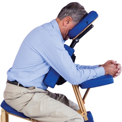
Posturing equipment
You might also consider hiring posturing equipment which supports your neck, back and shoulders. Speak to your ophthalmologist or contact MDFA for more information.

Preparing prior to surgery
Preparing before you go into hospital is important. If you need to posture, you’ll be expected to start as soon as you return home. Before you go into hospital consider things such as:
- doing laundry and housework, and ensuring the home is clear of trip hazards
- paying all household bills due during your recovery time
- shopping and food preparation (e.g. prepare meals ahead and freeze)
- arranging delivery of meals or other social services
- hiring posturing equipment, allowing a week or so for this to be delivered
- making sure your posturing equipment and aids are positioned where you want to sit
- arranging for someone to stay with you if you live alone.
While posturing you may wish to:
- keep the things you use frequently close by (for example, tissues, drinks, books, phone, tablets, laptop)
- drink through a straw instead of using a glass or cup
- use a laptop or a tablet such as an iPad, instead of watching TV.
Managing vision loss
Surgery is usually very successful at reattaching the retina. Unfortunately for some people, the operation may be successful at reattaching the retina but it may not bring back detailed central vision or areas of peripheral vision. This can happen in any circumstance. However, the risk is higher the longer the retina has been detached without any treatment.
However, if you do experience significant vision loss, a key priority is maintaining quality of life and independence.
Get the fact sheet
The information on this page is available as a printed fact sheet. You can preview or download the fact sheet below. Alternatively, you can order a free printed copy to be sent to you.
Download the publication today.
Download


